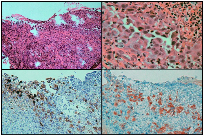
Figure 2.

An antral ulcer (upper left, H&E stain, 100X magnification), with large epithelioid tumor cells (upper right, H&E stain, 400X magnification). The tumor cells stain for cytokeratin 7 (left lower panel, immunostaining, 200 x magnification) and for BRAF V600E mutant (right lower panel, immunostaining, 200 x magnification) by immunohistochemistry
