
Figure 2.
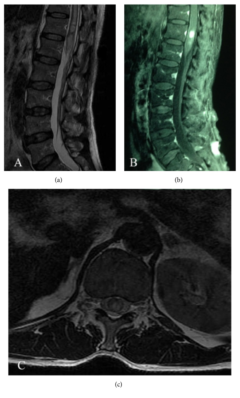
(a) Preoperative T2-weighted sagittal MRI demonstrating increased signal intensity. (b) Preoperative T1-weighted sagittal MRI revealing a well-defined tumor with significant enhancement after gadolinium injection. (c) Preoperative T1-weighted axial MRI showed well-circumscribed lesion inside the spinal cord at the T12 level.