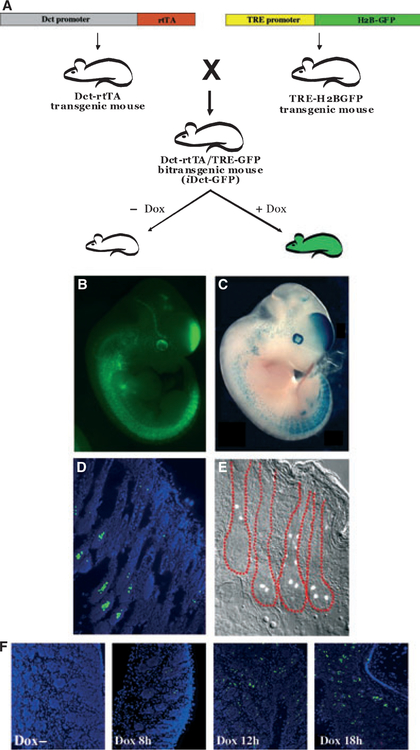
Figure 1.
(A) Making of iDct- green fluorescent protein (GFP) mice. Dct-rtTA transgenic mice were crossed with the TRE-H2BGFP mice to get the iDct-GFP bi-transgenic mice, which show tightly regulated doxycycline-inducible GFP expression. (B) An E11.5 iDct-GFP embryo. (C) A Dct-LacZ embryo (courtesy, Dr. Ian Jackson). (D) GFP expression in the skin of a 7 days old pup is detectable in the bulb and bulge regions of the hair follicles. Blue, DAPI. (E) Skin section from an adult mouse, showing GFP+ cells in the hair follicles (demarcated in red). (F) Induction of GFP expression by a single intraperitoneal injection of doxycycline. Images have been modified from Zaidi et al.

