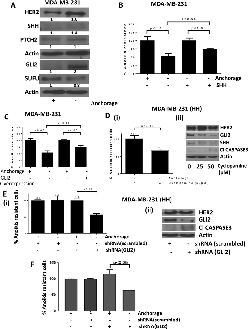
Figure 3– SHH signaling promote anoikis resistance.
A) Western blot analysis for protein modulations in adherent vs anchorage independent MDA-MB-231 cells. B) Anoikis assay for MDA-MB-231 cells treated with SHH ligand and respective control cells. C) Anoikis assay for MDA-MB-231 cells transfected with empty vector or GLI2 overexpression plasmid. D) Anoikis assay (i) and western blot analysis (ii) for HH cells treated with cyclopamine, a pharmacological inhibitor of SHH signaling. E) Anoikis assay (i) and western blot analysis (ii) for HH cells transfected with scrambled shRNA or GLI2 shRNA to compare anoikis resistance. F) Anoikis assay in SKBR3 cells transfected with GLI2 shRNA. Values were plotted as mean ± SD (n=3).

