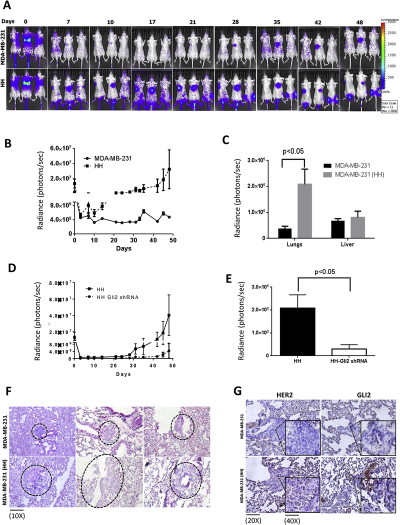
Figure 6– Increased metastasis of HH cells in vivo and increased expression of HER2 and GLI2 in tumors from anoikis resistant MDA-MB-231 and HH cells.
About 0.5 × 10⁶ MDA-MB-231 or HH anoikis resistant cells were injected by tail vein route in athymic nude female mice (n=6). The mice were imaged periodically for bioluminescence after luciferin injections. A&B) Time dependent luminescence imaging data from athymic nude mice for MDA-MB-231 and HH cells. C) Luminescence imaging data from isolated lungs and livers of from MDA-MB-231 and HH injected mice at the end of experiment. D) Time dependent luminescence signal from mice injected with HH and HH with GLI2shRNA cells. E) Average luminescence of the lungs from HH and HH-GLI2 shRNA group, which were imaged ex vivo at the end of the experiment. Values were plotted as means ± SEM (n=6). The lungs from mice of both the groups were fixed in formalin and processed for microscopic evaluation of tumors. F) H&E staining for tumors from different mouse lung samples of MDA-MB-231 and HH injected mice. G) immunohistochemical staining for HER2 and GLI2 in tumors from MDA-MB-231 and HH injected mice lungs. The inset in all panel shows enlarged view of tumor cells. Values were plotted as mean ± SEM (n=6).

