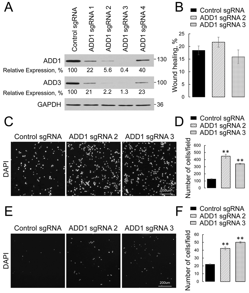
Figure 2: Loss of ADD1 increases transfilter migration and Matrigel invasion of lung cancer cells.
ADD1 expression was down-regulated H1573 cells using CRISPR/Cas9-mediated gene editing. (A) Immunoblotting analysis shows co-depletion of ADD1 and ADD3 by 4 different ADD1 small guide (sg) RNAs. (B) Quantification of the planar migration of the control and ADDl-depleted H1573 cell monolayers at 24 h post-wounding. (C, D) Representative images and quantitative analysis of the DAPI-labeled control and ADD 1-deficient HI 573 cells after 16 h of transfilter migration in the Boyden chamber. (E, F) Representative images and quantitative analysis of the DAPI-labeled control and ADD 1-deficient H1573 cells after 24 h invasion into Matrigel. Data are presented as mean ± SE (n =3); **p < 0.005, as compared to the control sgRNA-transfected group.

