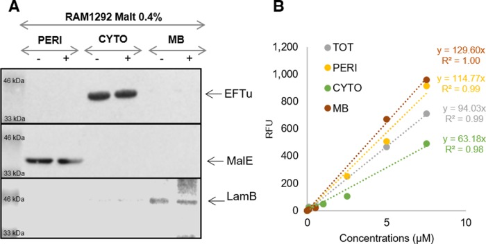
Figure S8. Distribution of specific proteins of various cell fractions and standard calibration curves of Cpd-1.
(A) Western blot to confirm the distribution of specific proteins of the different cellular fractions of RAM1292 induced with 0.4% maltose shown in Fig 5. Antibodies were directed against EfTu (1/30,000), MalE (1/5,000) and LamB (1/10,000) that are well-known specific proteins of the cytoplasm (CYTO), periplasm (PERI), and membrane (MB) fractions respectively. (B) Standard calibration curves of Cpd-1 fluorescence used to determine the number of accumulated molecules in the different compartments of the RAM1292 cells shown in Fig 5.

