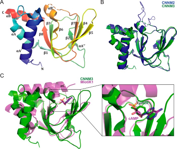
Figure 2.
Crystal structures of CNBH domain of CNNM proteins. A, cartoon representation of CNNM3 CNBH domain, colored blue (N terminus) to red (C terminus). A disordered loop of 31 amino acids is indicated by a dashed line. The CNNM3 CNBH domain structure shows the typical fold of a cyclic nucleotide–binding domain: a wide antiparallel β-roll capped by an α-helical bundle. B, overlay of cartoon representations of CNBH domains of CNNM2 (blue) and CNNM3 (green). C, structural overlay of the CNBH domain of CNNM3 (green) with the cyclic nucleotide–binding domain of the bacterial K+ channel from Mesorhizobium loti (magenta; PDB code 1VP6). The M. loti K+ channel has an additional C-terminal helix (αC) that contacts the bound cAMP ligand. In CNNM3, a tyrosine side chain blocks the nucleotide-binding site.

