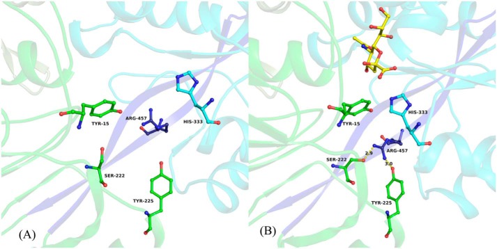
Figure 3.
Close-up view of hinge region and amino acids from the binding pocket of Hd-SatA. Neu5Ac and its interacting residues in the binding pocket of Hd-SatA are shown in a ball-and-stick model. A close-up view clearly shows the conformational changes in the amino acids before (A) and after (B) Neu5Ac binding. Neu5Ac is shown in B. The two β strands that form the hinge are shown in dark blue color. The figure was created using PyMOL (Schrödinger) (54).

