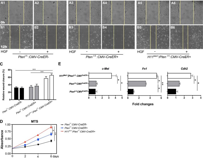
Figure 6.
The conditional expression of mouse Met transgene and homozygous Pten deletion increases cell proliferation and migration in MEFs. A and B, wound-healing assay of MEFs. Shown are before (A) and after (B) images of scratches in the monolayer of PtenL/L:CMVCreER-, PtenL/L:CMVCreER+, and H11Met/+/PtenL/L:CMVCreER+ MEFs cultured in the absence/presence of HGF for 12 h. C, graphical representation of relative wound closure. The data represent quantifications of multiple images from two independent experiments ± S.D. D, graphical representation of cellular proliferation of PtenL/L:CMVCreER−, PtenL/L:CMVCreER+, and H11Met/+/PtenL/L:CMVCreER+ MEFs measured by MTS reduction. The data represent the mean ± S.D. of three independent experiments. E, qRT-PCR analysis of Met, Fn1, and Cdh2 expression in PtenL/L:CMVCreER+ and H11Met/+/PtenL/L:CMVCreER+ MEFs. Bars, mean ± S.D. (error bars) of three independent experiments. *, p < 0.05; **, p < 0.01.

