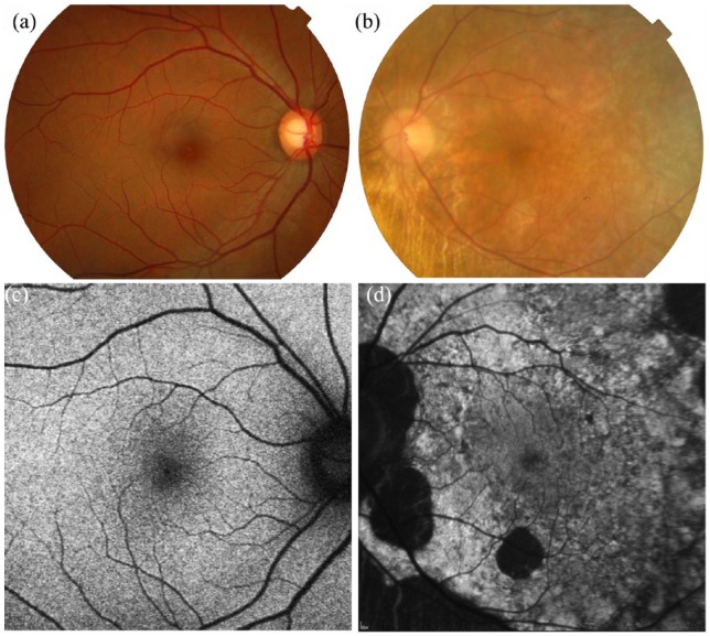
Figure 3.
Colour fundus photography and fundus autofluorescence of (a, c) an unaffected female subject versus (b, d) a CHM female carrier (c.715C>T, p.[Arg239*]). (b) Fundus photograph demonstrates peripapillary changes in CHM female carrier with accompanying peripheral pigmentary and atrophic areas similar to affected male patients. (d) Fundus autofluorescence shows a speckled pattern with intermixed areas of high- and low-density autofluorescence.

