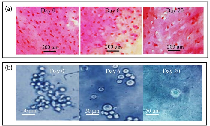
Figure 6.
(a): Safranin-o staining of printed chondrocyte-seeded constructs in 0, 6, and 20 days’ culture from the T-7 group. Note the increase in cell size and highly positive stain for safranin-o in day 20 culture as compared with 0 and 6 days’ cultures, (b): Photomicrographs of chondrocyte-seeded constructs at different days of culture from the T-7 group; Note the increase in cell size with the progress of culture, Toluidine blue staining.

