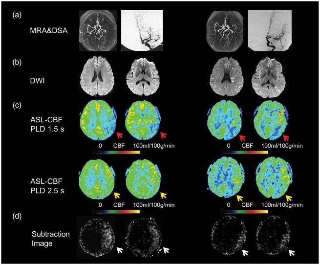
Figure 2.
Two delay ASL presentation of an asymptomatic patient (the first two columns) and symptomatic patient (the last two columns). Severe stenosis of left MCA was shown on MRA and DSA (a) in a 32-year-old asymptomatic patient and a 34-year-old symptomatic patient. Multiple DWI lesions located in cortical, subcortical regions and periventricular white matter were observed in the symptomatic patient (b). Decreased CBF was noted on ASL with PLD 1.5 s (C, red arrows) but normal CBF with PLD 2.5 s (C, yellow arrows) in the asymptomatic patient, whereas decreased CBF with PLD 2.5 s (C, yellow arrows) was still noted in the symptomatic patient. The total collateral perfusion area on the presented two slices was 65.5 cm2 and 32.7 cm2 on subtraction images between PLD 1.5 s and 2.5 s (D, white arrows), respectively. DWI: diffusion weighted imaging; CBF: cerebral blood flow; MCA: middle cerebral artery; PLD: post labeling delay.

