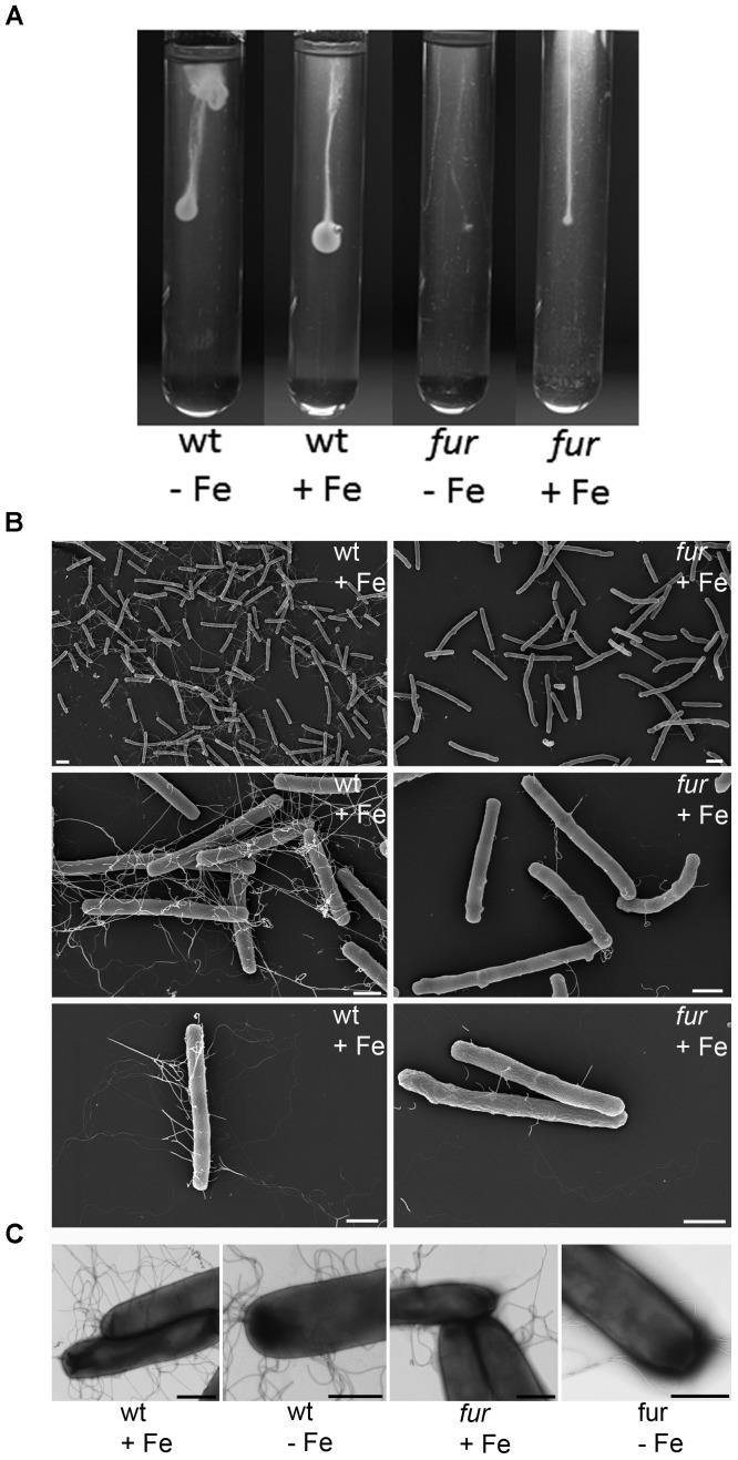
FIGURE 4.
Scanning/transmission electron microscopic images and motility assays of C. difficile wild type and the corresponding fur mutant. (A) CDMM agar filled glass tubes containing 0.2 μM (-Fe) and 15 μM (+Fe) iron sulfate were inoculated with wild type (wt) and the corresponding fur mutant strain and incubated anaerobically for 24 h. Scanning electron microscopy picture of C. difficile (B, right panel) and the fur (mutant B, left panel) grown in CDMM containing 0.2 μM (-Fe) and 15 μM (+Fe) iron sulfate are shown. Negative staining (C) also depicts less flagellation of the fur mutant and no detectable other appendage-like structures on the surface of C. difficile like pili or fimbriae. Bars represent 2 μm in (B), top 2 images and 1 μm in all other images.

