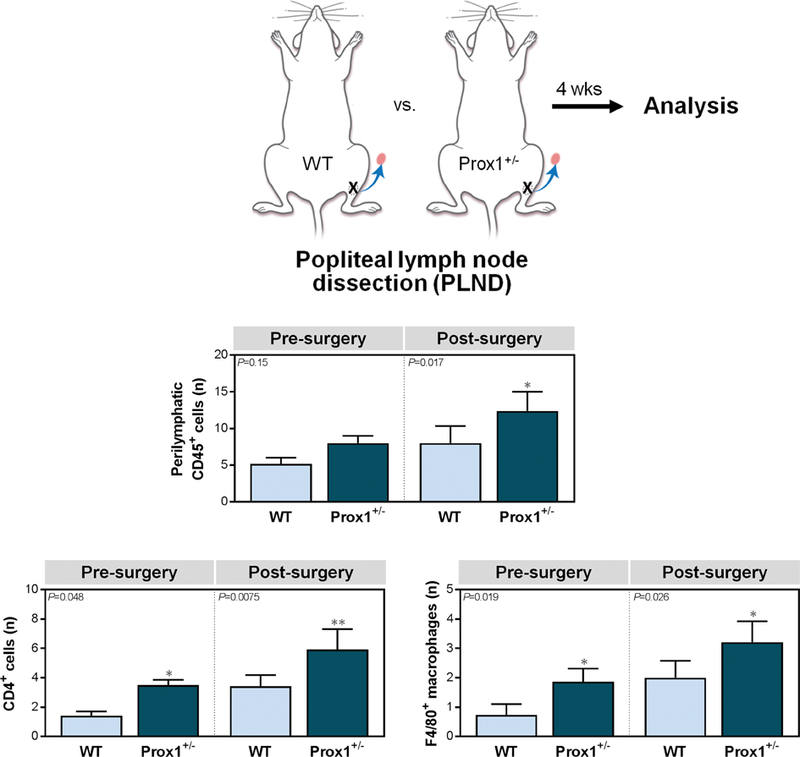
Figure 2. Prox1+/− mice have amplified inflammatory responses following lymphatic injury.

A. Schematic depiction of popliteal lymph node dissection (PLND).
B. Quantification of CD45+ cells within 50 μm of LYVE-1+ lymphatic vessels [one-way ANOVA, mean (S.D.); n=4–5/group; 4 high-powered fields (h.p.f.)/mouse, P=0.15 and P=0.017 for pre- and post-PLND, respectively].
C. Quantification of CD4+ cells [left; one-way ANOVA, mean (S.D.); n=4–5/group; 4 h.p.f./mouse, P=0.048 and P=0.0075 for pre- and post-surgery evaluation, respectively] and F4/80+ macrophages [right; one-way ANOVA, mean (S.D.); n=5/group; 4 h.p.f./mouse, P=0.019 and P=0.026 for pre- and post-PLND, respectively] within 50 μm of LYVE-1+ lymphatic vessels.
