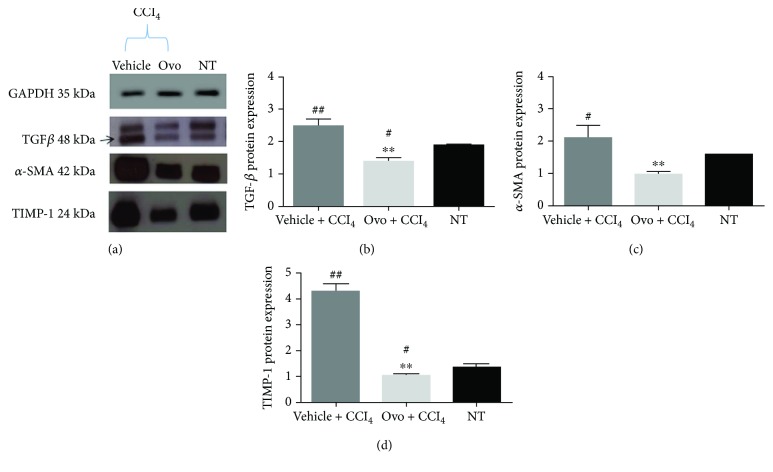
Figure 3.
Protein expression of liver fibrosis markers. (a) A representative experiment of Western blot analysis of cytosolic extracts obtained from hepatic tissues of mice treated with ovothiol A or vehicle, after induction of liver fibrosis, compared to samples of healthy mice (NT), using antibodies specific for TGF-β, α-SMA, and TIMP1. Histograms of the densitometry analysis of protein bands obtained by Western blot for liver markers: (b) TGF-β; (c) α-SMA; and (d) TIMP1. Data were normalized for GAPDH. Data are expressed as mean ± SD, n = 7. The significance was determined by ANOVA test. (#p < 0.05) and (##p < 0.01) represent significance compared to NT; (∗∗p < 0.01) represents significance compared to the treated with vehicle + CCl4.

