Figure 4.
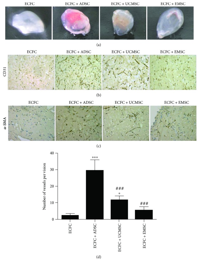
In vivo proangiogenic properties of ADSCs, UCMSCs, and EMSCs. (a) Macroscopic views of representative explants at day 7. Matrigel containing ECFCs with/without different MSCs was subcutaneously injected into the dorsal subcutaneous of nude mouse. The explants were excavated after 7 days. The group of ECFCs was used as control. (b) Human CD31 staining confirmed blood vessel structure and endothelial cell origin. Positive staining was shown in brown. The group of ECFCs was used as control. (c) Human α-SMA staining identifies human mesenchymal stromal cells within the implant. α-SMA positive staining was shown in brown. α-SMA, alpha-smooth muscle actin. (d) Statistical analysis of microvessel number in matrigel explants. Microvessel number was determined by counting the luminal structure containing erythrocytes in each field. Bars represent the mean value ± SD. ∗∗∗ P < 0.001, ∗ P < 0.05, compared with ECFC group; ###P < 0.001, compared with ECFC + ADSC group. N = 3 separate experiments.

