
Figure 1.
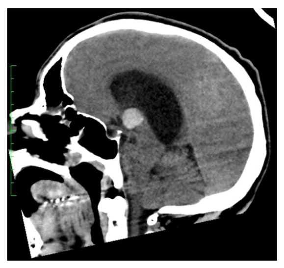
The marked lateral ventricle hydrocephalus secondary to a 16 x 15 x 15 mm colloid cyst within the third ventricle seen on CT scan at admission from the sagittal view.
Official websites use .gov
A
.gov website belongs to an official
government organization in the United States.
Secure .gov websites use HTTPS
A lock (
) or https:// means you've safely
connected to the .gov website. Share sensitive
information only on official, secure websites.
The marked lateral ventricle hydrocephalus secondary to a 16 x 15 x 15 mm colloid cyst within the third ventricle seen on CT scan at admission from the sagittal view.