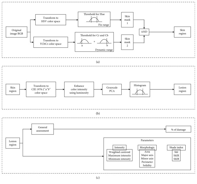
Figure 3.
Three-stage algorithm: (a) Stage 1—processing from skin image to segment a skin region using the AND of two skin masks (via HSV and YCbCr color space transformation). (b) Stage 2—lesion region segmentation from the skin region by means of CIE 1976 L∗a∗b∗ transformation followed by luminosity enhancement and PCA grayscale transformation. (c) Stage 3—Characterization of the lesion region. Calculation of the parameters for intensity, morphologic and the shade indices for macules.

