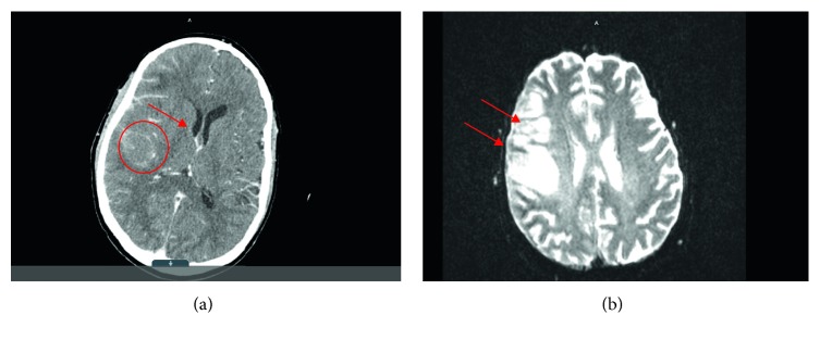
Figure 1.
(a) Initial CT scan showing increased diffuse enhancement of right cerebrum as well as ill-defined hypodensity in the posterior right frontal lobe (circle). Slight mass effect is shown upon right lateral ventricle with edema (arrow). (b) MRI brain with gadolinium showing parenchymal and leptomeningeal enhancement over the right frontal and temporal lobes with associated T2/FLAIR signal involving right frontal lobe cortex (arrows), finding suggestive of meningoencephalitis.

