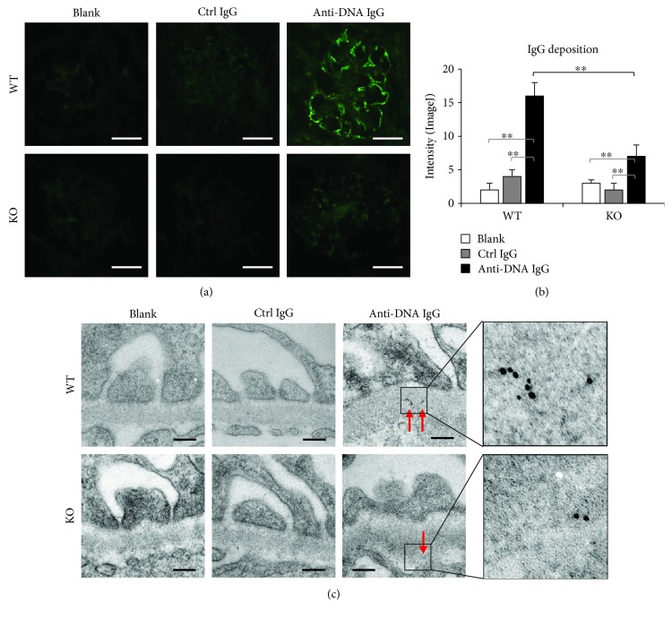
Figure 2.
Glomerular IgG deposition is attenuated in Fn14-deficient SCID mice. (a) IgG deposition was detected in glomeruli by immunofluorescence. Scale bar = 50 μm. (b) Fluorescent intensities of glomeruli were quantitated by ImageJ software. (c) By immunogold staining and transmission electron microscopy, IgG deposits were detected in glomeruli. Gold particles were indicated by red arrows. Scale bar = 200 nm. There were eight mice in each group. Representative images are shown. ∗∗ p < 0.01.

