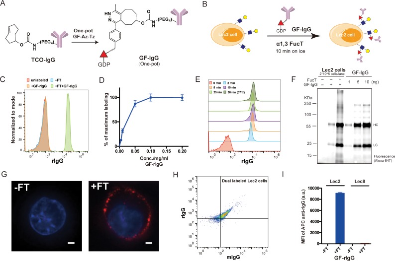
Figure 2.
Enzymatic transfer of IgG to the surfaces of Lec2 CHO cells: (A) The synthesis of a GDP-Fucose-conjugated IgG (GF-IgG). (B) Workflow of the FucT-catalyzed transfer of GF-IgG to the surface of Lec2 CHO cells. (C) Flow cytometry analysis of Lec2 cells treated with the enzyme FucT, the substrate GF-rIgG, or both. (D) Titration of GF-rIgG, concentrations ranging from 0.005 to 0.2 mg/mL in the reaction buffer; each reaction used 60 mU FucT and proceeded at room temperature for 30 min; mean ± SD (error bars), representative graph from three independent experiments. (E) Time course of enzymatic transfer of GF-rIgG to Lec2 cells on ice; reaction at 37 °C was used as the maximum labeling control. (F) Fluorescent gel imaging for detecting and quantifying rIgG (Alexa Fluor 647-labeled) molecules conjugated on the Lec2 cell surface. (G) Confocal microscopy images of Lec2 cells treated with or without FucT when incubated with Alexa Fluor 647-labeled GF-rIgG; nuclei were stained with Hoechst 33342. Scale bar: 2 μm. (H) Flow cytometry analysis of Lec2 cells simultaneously labeled with rIgG and mIgG. (I) Lec8 CHO cells without LacNAc expression were compared with Lec2 cells in the enzymatic IgG transfer as a negative control; mean ± SD (error bars), representative graph from three independent experiments. α1,3FucT is abbreviated as FT in all figures.

