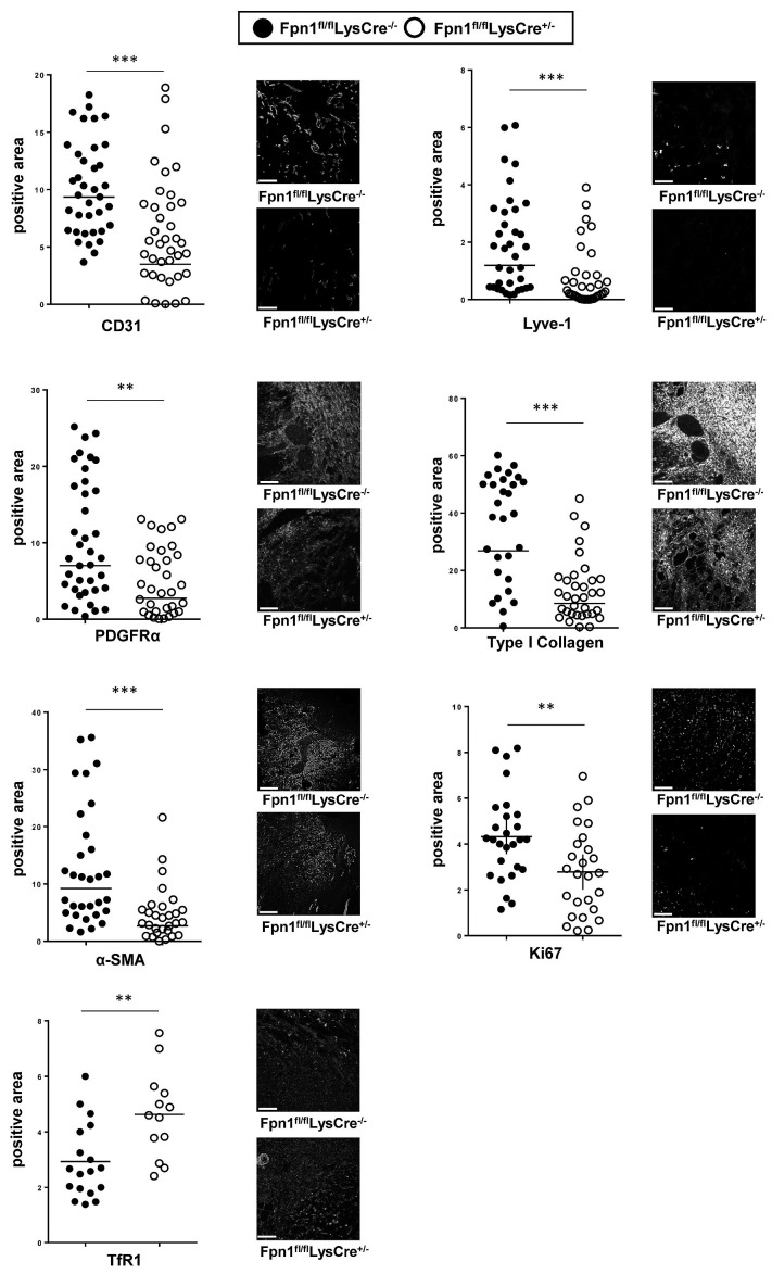
Figure 6.
Vessel and stromal cell reduction accompanied by iron deficiency and decreased proliferation in wounds of Fpn1fl/flLysCre+/− mice. Expression of CD31, Lyve-1, collagen-1, PDFGR, αSMA, Ki67 and TfR1 after skin wounding at 7 dpi was assessed by confocal microscopy and the positive area expressed as %. Each circle represents an analysis from a single confocal image (5-9 fields of vision/mouse, 6 mice/group), ***P<0.0001, **P<0.001. Representative confocal microscopy images are shown. Bars: 100 mm. Magnification: 40X.

