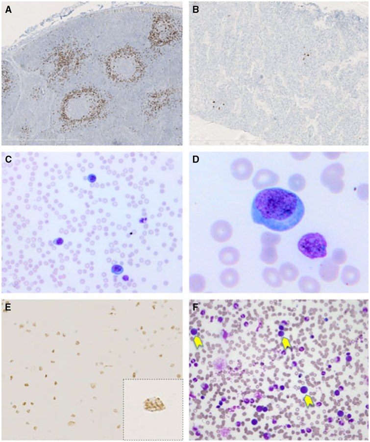
Figure 1.
Multicentric Castleman disease and HHV-8-positive polyclonal IgMλ B-cell lymphocytosis (patient 3). The two lymph node biopsies (before and after treatment) show typical features of multicentric Castleman disease. Immunostaining with the HHV-8 LNA-1-specific monoclonal antibody shows numerous, partly coalescent HHV-8-positive plasmablastic cells in the initial biopsy (A) and only a few HHV-8-positive cells in the second post-treatment biopsy (B). The blood smear shows numerous plasmablasts (C,D); the circulating plasmablastic cells stain positive for HHV-8 with the typical nuclear dotlike staining in brown (E). Bone marrow smear reveals the presence of large plasmablastic cells (yellow arrows) (F).

