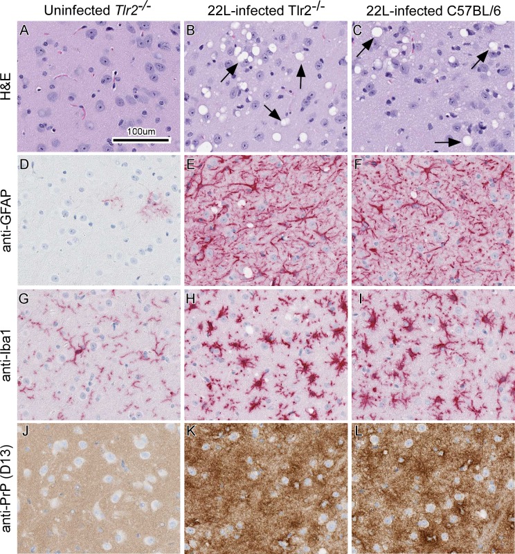
Fig 3. Representative neuropathology and immunohistochemical assessment of gliosis and PrP deposition in thalamic brain sections from uninfected and 22L-infected Tlr2-/- mice compared to 22L-infected C57BL/6 controls.
Tlr2-/- mice were uninfected (panels A, D, G, and J) or infected with scrapie strain 22L (panels B, E, H, and K). For comparison C57BL/6 mice were also infected with strain 22L (panels C, F, I, and L). Sections of thalamus were stained by H&E (A-C) or probed with antibodies that recognize GFAP (D—F), Iba1 (G—I), and PrP (J—L). Representative images of the thalamus are shown for all at the same scale as indicated in panel A. Black arrows in panels B and C indicate vacuoles present in the neuropil of 22L-infected mice. Similar histopathological results were seen when 22L-infected C3ar1-/- and C5ar1-/- mice were compared to infected Balb/c control mice.

