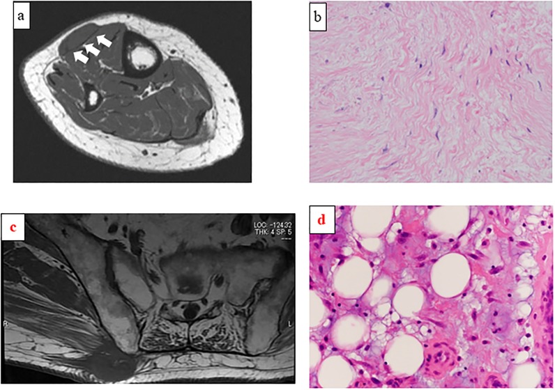
Fig 2. Magnetic resonance imaging findings of a superficial mass in the anterior part of the right lower leg in a 78-year-old woman.
Axial T1-weighted image showing a well-defined lesion with wider contact with the fascia at obtuse angles (a; Group 4 per the Galant classification). Pathologic examination of the resected specimen confirmed low-grade fibromyxoid sarcoma (b; hematoxylin-eosin staining; magnification ×200).

