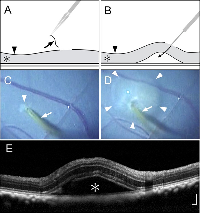
Fig 1. Local removal of internal limiting membrane and subretinal injection of balanced salt solution.
(A) Schematic drawing of local internal limiting membrane (ILM) removal showing the removed ILM (arrow), retina (asterisk), and intact ILM (arrowhead). (B) Schematic drawing of the subretinal injection procedure. Balanced salt solution (BSS) was injected with a 38-gauge cannula by placing the cannula tip in contact with the retinal nerve fiber layer. The arrow indicates the flow of the injected BSS; the asterisk and arrowhead indicate the retina and ILM, respectively. (C) Surgical photograph after local ILM removal. The arrow indicates the 38-gauge cannula; the arrowhead indicates the area of peeled ILM. (D) Surgical photograph during subretinal injection. The arrow indicates the 38-gauge cannula; arrowheads indicate the area of retinal detachment due to subretinal injection of BSS. (E) Optical coherence tomography 30 minutes after subretinal injection. The asterisk indicates retinal detachment caused by the procedure. Scale bar = 200 μm.

