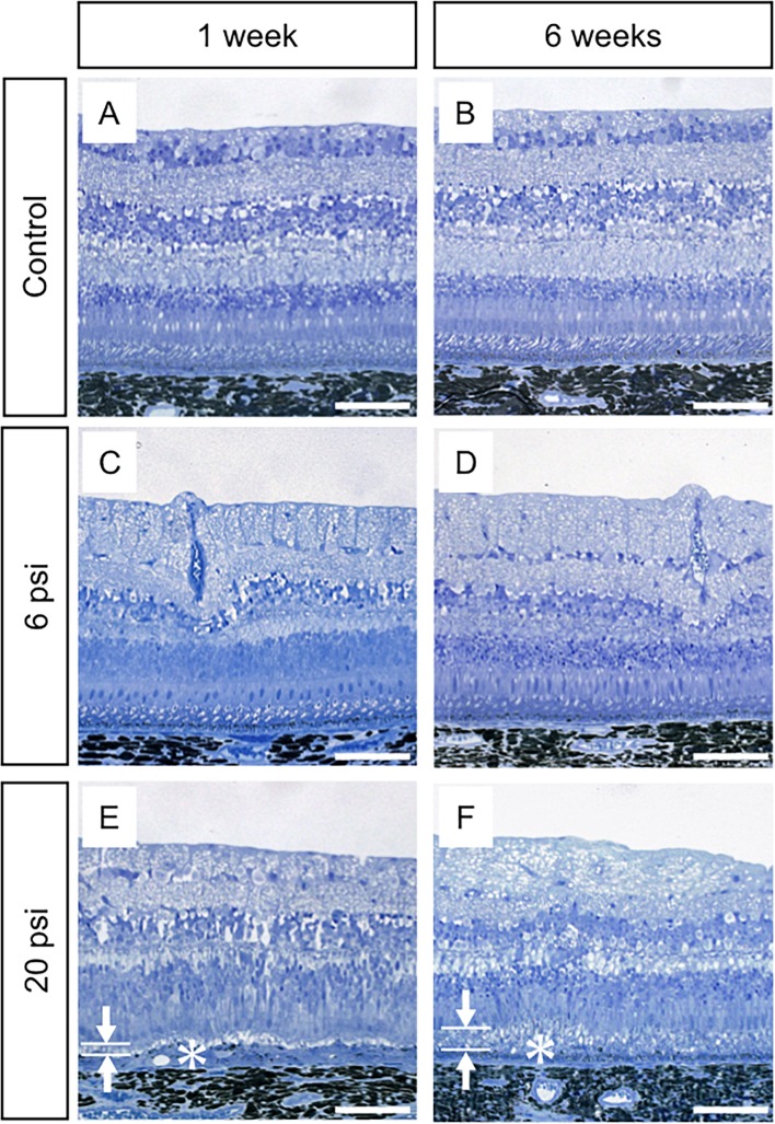
Fig 3. Light microscopy images of monkey retina after subretinal injection of balanced salt solution.
The retinal structures of both the control (no injection of balanced salt solution; BSS) and the minimum-pressure (BSS injection at 6 psi) groups are well-preserved at 1 week (A and C) and 6 weeks (B and D) after injection. The high injection pressure group (BSS injection at 20 psi) shows thinning of the photoreceptor outer segment layer (OS, arrows in E) and thickening of the retinal pigment epithelium (RPE) layer (asterisk in E) at 1 week after injection (E), while the photoreceptor cells are well-preserved. At 6 weeks after injection, the high-pressure group shows restoration of the OS (arrows in F) and flattening of the RPE (asterisk in F). Scale bars = 100 μm.

