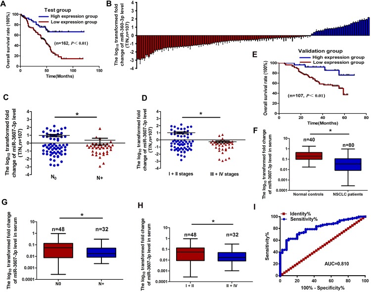
Fig 2. The relationship between miR-3607-3p expression levels in NSCLC tissues or serum and clinical significances.
(A) Kaplan-Meier overall survival curves according to high and low miR-3607-3p expression in 162 patients with NSCLC. (B) Quantitation of miR-3607-3p was performed using qRT-PCR in 107 NSCLC (T) and adjacent normal (N) tissues. The fold changes were calculated by relative quantification (2-ΔCt, U6 as the internal control). (C-D) miR-3607-3p expression was detected in primary tumor tissues and the patients were grouped according to lymph node metastasis status (C) or clinical stages (D). (E) Kaplan-Meier curves depicting overall survival according to the expression of miR-3607-3p from the validation set. (F) The expression levels of serum miR-3607-3p in 80 NSCLC patients and 40 healthy controls were measured by qRT-PCR and normalized to U6. (G-H) Serum miR-3607-3p expression was low in patients with NSCLC, indicated by lymph node metastasis (G) and different clinical stages (H). (I) Receiver operating characteristic (ROC) curve analysis of the miR-3607-3p assay ratio for detecting NSCLC patients. *, P < 0.05.

