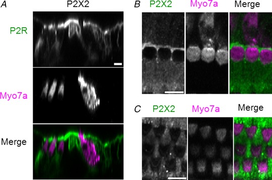
Figure 3. P2X2 receptor expression in the adult organ of Corti.

A, orthogonal projection of P2X2 receptor expression. P2X2 receptor immunofluorescence staining (green), myosin 7a (pink) and merged image. The hair cell cytoplasm is identified using myosin 7a as the label. B, surface view of P2X2 receptor immunohistochemistry in the IHC region of the adult mouse organ of Corti. C, P2X2 receptor staining in the OHC and Deiters’ cell region. Maximum projection image. Scale bar in all images = 10 μm.
