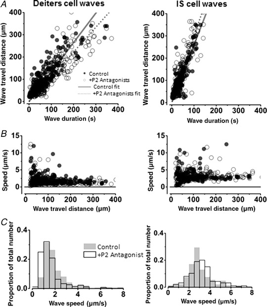
Figure 6. Slow Ca2+ waves are not blocked by P2 receptor antagonists.

A and B, Ca2+ wave propagation distance (A) and wave speeds (B) for Deiters’ and IS cell regions in 1.3 mm external Ca2+. Left‐hand column, Deiters’ cell waves; right‐hand column, IS cell waves. Open circles show wave propagation data in the presence of the P2 receptor antagonists PPADS and suramin. Closed circles show wave data under control conditions with no P2 receptor antagonists. The lines show the fit to the complete data sets: the dotted line shows the fit to the data in the presence of P2 receptor antagonists; the continuous line shows the fit to the control data in the absence of antagonists. C, frequency histograms (wave speed) in the presence of (shaded) and absence of (white) P2 antagonists. The slow Ca2+ waves in the inner sulcus region propagate faster than slow Ca2+ waves in the Deiters’ cell region. Addition of P2 receptor antagonists did not affect wave propagation speed or the distance the wave travelled before extinction in either of the regions.
