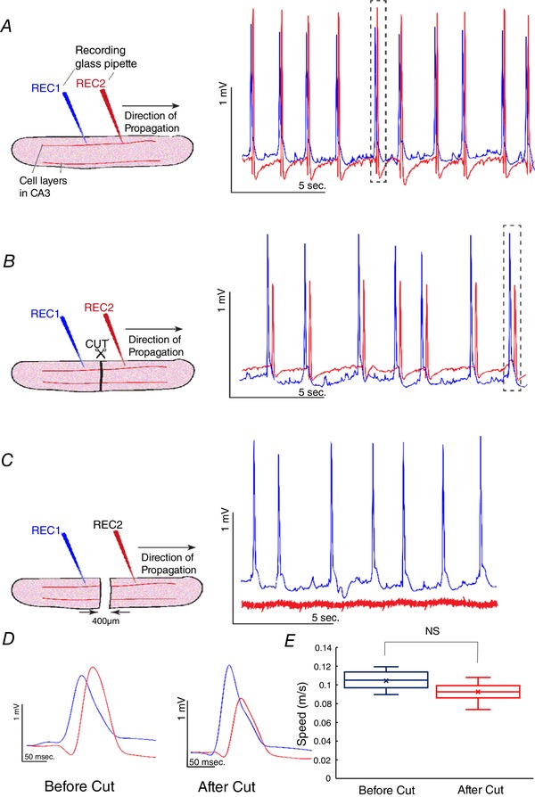
Figure 4. Slow hippocampal periodic activity propagation along the tissue with a complete cut in the hippocampal slice in vitro .

A, slow hippocampal periodic activity propagated from recording electrode 1 (REC1) to recording electrode 2 (REC2) at a speed of 0.10 ± 0.01 m s−1 before the cut. B, a complete cut in the hippocampal slice. Slow hippocampal periodic activity was observed to be propagating along the slice with a cut from REC1 to REC2 with speed similar to that recorded in an intact slice by activation of the neurons of the other side of the cut. C, slow hippocampal periodic activity stopped propagating when the gap was 400 μm. D, expanded windows of the single event of the slow hippocampal periodic activity before and after the cut revealing similar delays between two recording electrodes. E, speeds of slow hippocampal periodic activity before and after the cut. There is no significant difference between the two speeds. NS, not significant. [Color figure can be viewed at wileyonlinelibrary.com]
