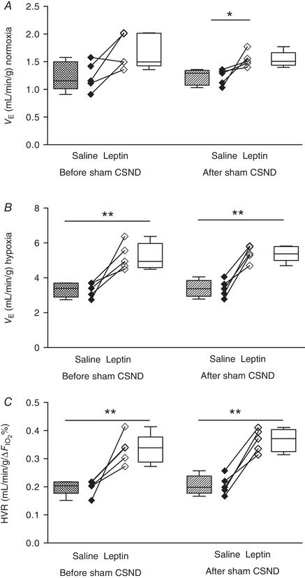
Figure 6. Leptin augmented minute ventilation (V E) and the hypoxic ventilatory response (HVR) and this effect was not modified by sham carotid sinus nerve dissection (CSND) in C57BL/6J mice during quiet wakefulness.

A and B, V E was calculated as tidal volume per body weight (V T) × respiratory rate (RR) at 21% O2 (A) and 10% O2 (B) during saline and leptin continuous infusion (120 μg/day, s.c.) measured before (n = 5) and after (n = 5) sham CSND. C, HVR represents the ratio of a change in V E to a change in the fraction of inspired oxygen (HVR = ΔV E/Δ). * P < 0.05, ** P < 0.01 using the Mann–Whitney test for unpaired comparisons.
