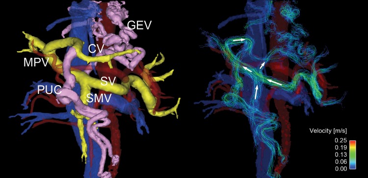
Figure 4:
Oblique frontal view four-dimensional (4D) flow MR images in a 54-year-old woman with high-risk varices. Radial 4D flow MRI can depict both segmented anatomic images (left) and streamline reconstruction (right). Portal system is colored yellow, and abnormal collaterals (varices) are pink. In this patient, hepatofugal flow is observed in a large periumbilical collateral (PUC) arising from the left portal vein, and an enlarged coronary vein (CV) that supplies flow to prominent gastroesophageal varices (GEV). Hepatopetal flows are observed in splenic vein (SV) and superior mesenteric vein (SMV). In this patient, the fractional flow change in the main portal vein (MPV) was −0.14, which helped to predict the presence of high-risk varices that were observed at endoscopy.

