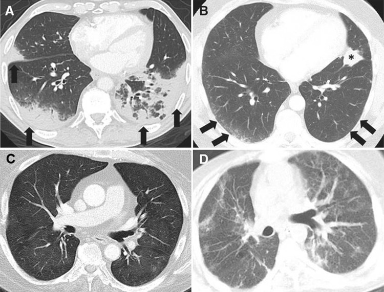
Figure 6:
Axial chest CT images show the spectrum of radiographic patterns of immune-checkpoint inhibitor–related pneumonitis, which includes, A, the cryptogenic organizing pneumonia (COP) pattern, B, the nonspecific interstitial pneumonia (NSIP) pattern, C, the hypersensitivity pneumonitis (HP) pattern, and, D, the acute interstitial pneumonia (AIP)/acute respiratory distress syndrome (ARDS) pattern. A, The COP pattern is characterized by multifocal bilateral parenchymal consolidations with peripheral and lower lung distribution, with ground-glass opacities (GGOs) and reticular opacities (arrows). B, The NSIP pattern demonstrates GGOs and reticular opacities predominantly in a peripheral and lower lung distribution (arrows). * = Lung tumor burden. C, The HP pattern demonstrates diffuse GGOs and centrilobular nodularities, with scattered areas of air trapping. D, The AIP/ARDS pattern is characterized by diffuse or multifocal GGOs or consolidations, along with lung volume loss and traction bronchiectasis. (Reprinted, with permission, from reference 66).

