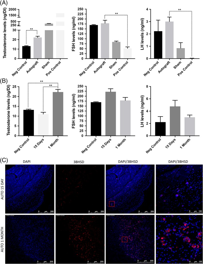
Figure 3.

(A): Leydig stem cells function was validated by comparing the production of testosterone, follicle‐stimulating hormone, and luteinizing hormone in negative control (castrated), autograft, sham, and positive control (testopel) using enzyme‐linked immunosorbent assay. (B): To further confirm that it is the stem Leydig cells that undergo differentiation in the grafts, hormone levels were compared after 15 days and 1‐month autograft. (C): Immunostaining showing 3BHSD expressing cells in autografts from 15 days and 1‐month autograft. Significance of differences was calculated using t test; **, p < .01; *, p < .05, and ns (non‐significant) (p > .05). Abbreviations: BHSD, beta hydroxysteroid dehydrogenase; DAPI, 4′,6‐diamidino‐2‐phenylindole; FSH, follicle‐stimulating hormone; LH, luteinizing hormone.
