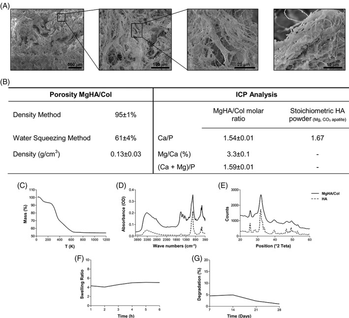
Figure 3.

Characterization of 3D biomimetic MgHA/Col scaffold. (A) Scanning Electron Microscopy micrographs at different magnifications, showing scaffold porosity and micro‐architectures of the hybrid composite. (B) Porosity and chemical composition of the hybrid MgHA/Col scaffold. (C) TGA, (D) FTIR, and (E) XRD analysis. In plots D and E, the solid line indicates the MgHA/Col, while the dashed one the HA. (F) Evaluation of the swelling ratio and (G) percentage of degradation of MgHA/Col scaffolds overtime.
