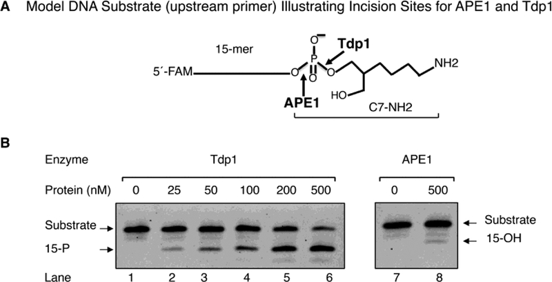
Fig. 4.
Comparison of Tdp1 and APE1 activities on the model DNA substrate. (A) Schematic of the model DNA substrate. This substrate mimics post-proteasomal treatment of a PARP-1 DPC. The 15-mer strand DNA has FAM at the 5´-end. (B) Representative phosphorimage of incision activities of Tdp1 (lanes, 1–6) and APE1 (lanes, 7–8). The incision reactions were performed as described under “Materials and Methods.” Reactions were initiated by the addition of different concentrations of Tdp1 (25 to 500 nM, lanes 2–6) or APE1 (500 nM, lane 8). Incubation was 20 min at 37 °C. Representative results from three independent experiments are shown. The migration positions of the substrate and the products with 3´-PO4 (15-P) and 3´-OH (15-OH), respectively, are indicated.

