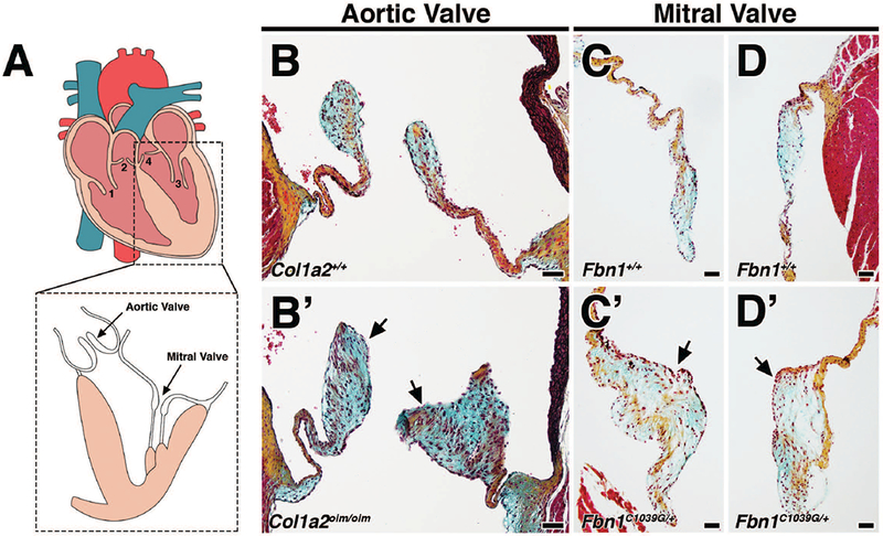
FIGURE 1: Mouse models of myxomatous valve disease.

A: The four heart valves consist of the tricuspid valve (1), pulmonic valve (2), mitral valve (3), and aortic valve (4). B-D: Movat’s pentachrome staining of aortic and mitral valves in 4 month-old Col1a2+/+;Fbn1+/+ wildtype mice depict collagen (yellow), proteoglycan (blue), and elastin (black). Myocardium is stained red. B’-D’: Age-matched 4 month-old Col1a2oim/oim and Fbn1C1039G/+ mutant mice exhibit myxomatous aortic and mitral valves, respectively. Myxomatous valves exhibit significant thickening resulting from increased proteoglycan deposition (arrows, blue) and abnormal ECM organization. Scale Bar = 50μm.
