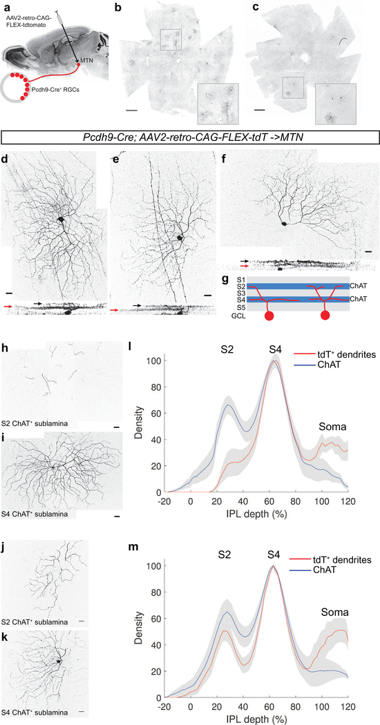
Figure 3: Dendritic architecture of Pcdh9-Cre+ RGCs.
(a) Schematic of retrograde infection with AAV2-retro. (b,c) En face view of retinal whole mounts of Pcdh9-Cre animals injected with AAV2-retro into the MTN. Many tdTomato+ RGCs are found throughout the retinas. Insets show higher magnification view of selected regions. (d-f) Individually labeled Pcdh9- Cre+ RGCs retrogradely labeled from the MTN with AAV2-retro-CAG-FLEX-tdTomato. Horizontal rotations of the confocal image stacks are shown below. 9 of 17 cells exhibit a primarily S4 dendritic lamination (example in d), and 8/17 are more bistratified (examples in e, f). Black and red arrows denote lamination with the ChAT+ plexuses in the S2 and S4 sublaminae, respectively. (g) Schematic of sublaminae in the inner plexiform layer showing locations of ChAT plexuses and Pcdh9-Cre+ RGC dendrites (h-k) Dendrites of retrogradely labeled Pcdh9-Cre+ RGCs cofasciculating with S2 (h, j) and S4 (I, k) ChAT plexuses for exemplary monostratified (d) and bistratified (e, f) cells. (l, m) Dendritic stratification analysis of monostratifed (l) and bistratified (m) cells dendrites compared with position of ChAT as a function of IPL depth. Red and blue lines denote mean with grey showing ±SEM, n=9 monostratified, n=8 bistratified. Scale bars: b,c, 500 microns, d-k, 25 microns.

