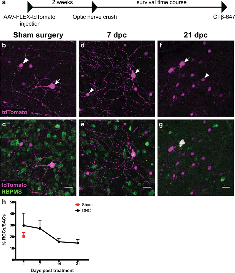
Figure 5: Pcdh9-Cre+ RGCs can survive up to 21 days after optic nerve injury.
(a) Timeline of optic nerve crush experiments. (b-f) Representative images of Pcdh9-Cre+ retinas infected with AAV2-CAG-FLEX-tdTomato (magenta) and labeled with anti-RBPMS antibody (green). White arrows mark RBPMS-positive RGCs and white arrowheads mark RBPMS-negative starburst amacrine cells (SACs). (b-c) In the sham surgery condition, the percentage of infected RGCs to SACs is 20.3%, and the RGCs have dendritic arbors (n=2 retinas). (d-e) This percentage at 7 dpc is similar to the sham condition and dendritic arbors are clearly present (n=2 retinas). (f-g) By 21 dpc, the percentage of infected RGCs to SACs is 14.5% and dendrites are absent (n=3 retinas). (h) Percentage of infected RGCs to SACs at different time points following optic nerve crush (Black circles) or from Sham treated animals (red square). The number of Pcdh9-Cre+ RGCs relative to Pcdh9-Cre+ SACs decreases around 14 dpc but shows no further reduction by 21 dpc; these differences are not statistically significant (One-way ANOVA, p = 0.2081). Error bars denote mean and SEM. Numbers of retinas and RGCs examined for each time point were: Sham: N=2 retinas, 104 RGCs; 1 dpc: N=3 retinas, 136 RGCs; 7 dpc: 2 retinas, 69 RGCs; 14 dpc: 4 retinas, 129 RGCs; 21 dpc: 3 retinas, 128 RGCs. Scale bars: b-g, 20 microns.

