Fig. 1.
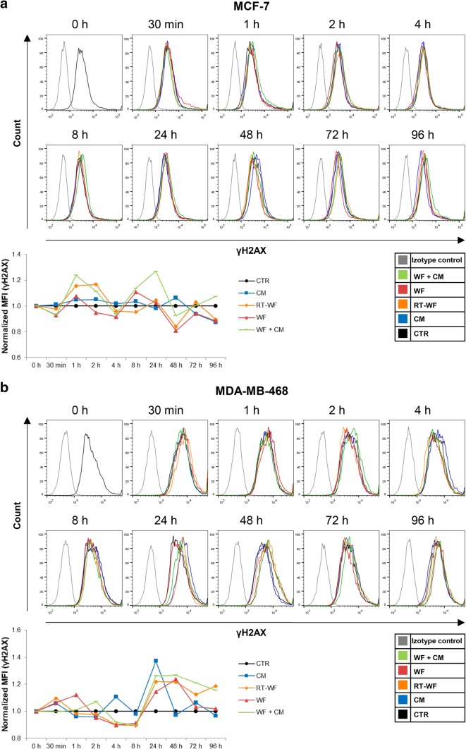
IORT alters the level of wound fluid-induced double-strand breaks (DSB) in breast cancer cells. Figure presents time course of the γH2AX level changes measured by flow cytometry in the a MCF-7 cells and b MDA-MB-468 cells. Data are presented as histograms from representative samples and as graphs of mean fluorescence intensity (MFI) normalized to the untreated control (mean of n experiments ± standard deviation). N = at least 3 independent biological replicates. CM conditioned medium collected from irradiated cells, RT-WF cells stimulated with 10% wound fluid collected after surgery and intraoperative radiotherapy, WF cells stimulated with 10% wound fluid collected after surgical excision, WF + CM cells stimulated with 5% conditioned medium and 5% surgical wound fluid

