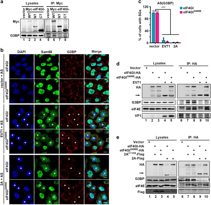
Fig. 6. EV71 and 2A block tSG formation by disrupting eIF4GI-G3BP interaction.
a HeLa cells were transfected as indicated. Cell lysates were subjected to IP with an anti-Myc antibody. b eIF4GI-HA- or eIF4GIG689E-HA-HeLa cells were transfected with vector or 2A for 24h, or infected with EV71 (MOI = 10) for 5 h, then treated with 200 μM AS for 1 h, and stained with Sam68 (green) and G3BP (red). Sam68 served as an indicator of EV71 infection or 2A expression. “ + ” indicates EV71-infected or 2A-expressing cells. c Quantitation analysis of vector-transfected, EV71-infected, or 2A-expressing cells with tSGs in (b). n = 3, 240 cells/condition were counted, mean ± SD; ***p < 0.001. d eIF4GI-HA- or eIF4GIG689E-HA-HeLa cells were infected with EV71 as indicated, and cell lysates were subjected to IP with an anti-HA antibody. The bound proteins were analyzed via WB. VP1 indicated EV71 infection. Arrow indicates eIF4GI cleavage products. e eIF4GI-HA- or eIF4GIG689E-HA-HeLa cells were transfected with Flag-tagged 2A or 2AC110S for 24 h and analyzed as in (d). Scale bars, 10 μm

