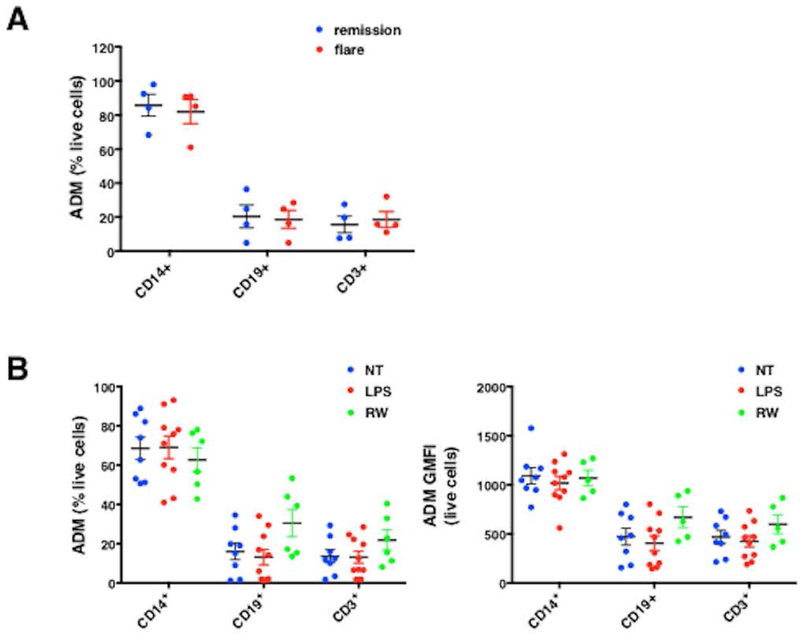
Figure 5. Monocytes are a major source of ADM within PBMCs.

(A-B) Intracellular ADM expression in PBMCs from SCLS patients obtained during flares or remissions (A) or from controls left untreated (no treatment, NT) or stimulated with LPS (100 ng/ml) or 10% conditioned macrophage medium (“RW”) for 24 hours was evaluated by flow cytometry in the presence of monensin (B). Frequency of ADM producing cells is expressed as percentage of total live cells (A, B left panel), or geometric mean of fluorescence intensity of ADM expression/per cell (GMFI, B right panel).
