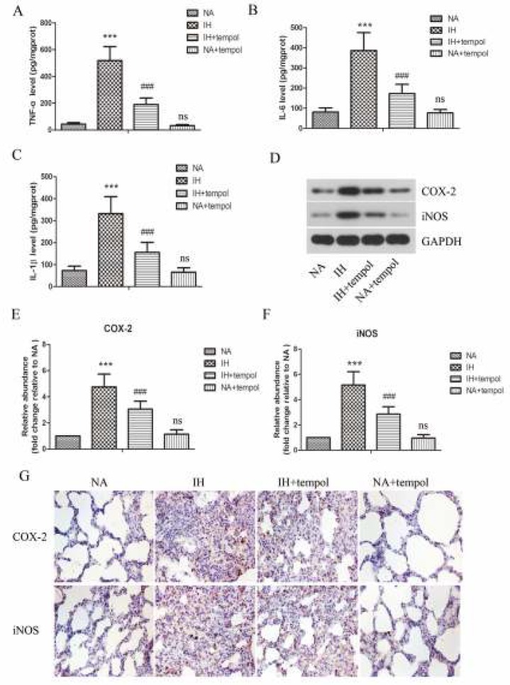
Figure 2.
Tempol restrained intermittent hypoxia-induced inflammatory response in lung tissues. The levels of TNF-α (A), IL-6 (B), and IL-1β (C) in lung tissues were assessed by ELISA. (D) The protein levels of COX-2 and iNOS were determined by Western blot assay. GAPDH was used as a loading control. (E-F) The gray-scale value of the bands was quantitatively analyzed. (G) The expressions of COX-2 and iNOS in lung tissues were evaluated by immunohistochemical staining. The experimental data were expressed as mean±SD (n=6). *** P<0.001, versus the NA group. ### P<0.001, versus the IH group. ns, no significance, versus the NA group

