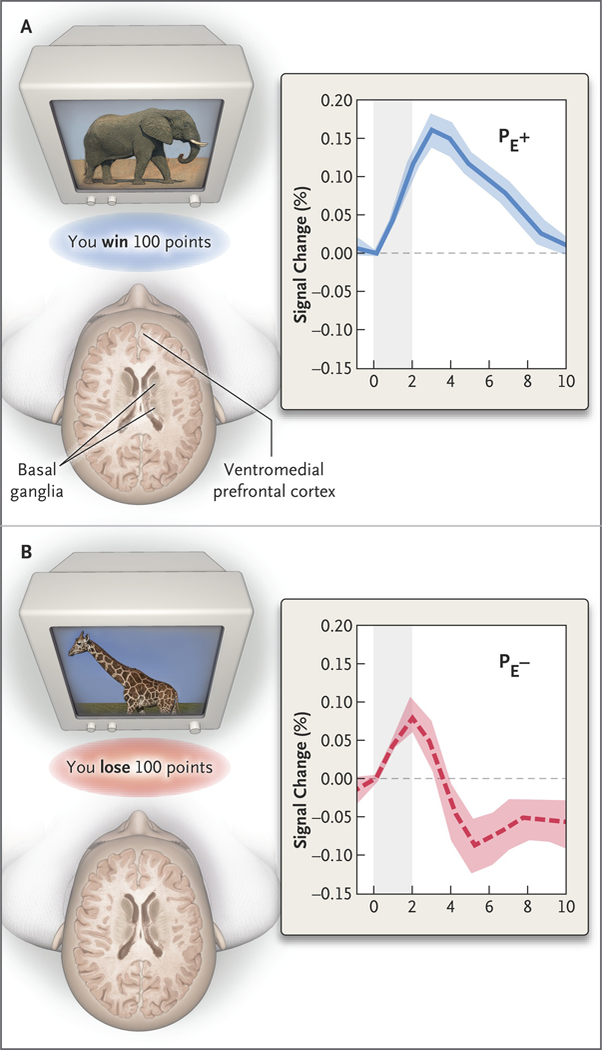
Figure 4. Format of a Functional MRI Study of Decision Making and Brain-Activation Data as They Relate to Prediction-Error Signals in the Basal Ganglia.

In this type of study, a research participant must learn to select one set of stimuli (e.g., the elephant) by pressing a button and to avoid another set of stimuli (e.g., the giraffe) by not pressing the button. Receipt of an unexpected reward elicits a positive prediction error (PE+), whereas failure to receive an expected reward, or a loss that is greater than expected, elicits a negative prediction error (PE−). The plots show responses to feedback in the basal ganglia during blood oxygen level–dependent functional MRI; the ventromedial prefrontal cortex is also shown. The timing of feedback is represented by the shaded column beginning at time zero. A PE+ occurs when the participant wins more than expected after pushing a button at the sight of the elephant. A PE− occurs when the participant loses more than expected after pushing a button at the sight of the giraffe. These positive and negative prediction errors generate the depicted reaction in the basal ganglia.
