
FIGURE 4.
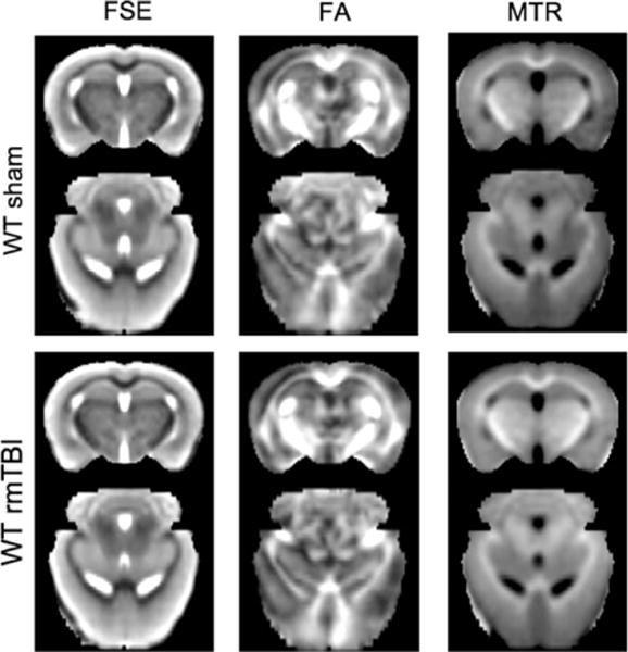
Comparisons of group-averaged brain data between sham-injured (top) and repetitive mild traumatic brain injury (rmTBI)-injured (bottom) mice (n = 4/group) using fast spin-echo (FSE) images, fractional anisotropy (FA) computed from diffusion tensor imaging, and magnetization transfer ratio (MTR). Group averages were obtained at a coronal slice 2mm posterior to bregma and an axial level 1.75mm ventral to bregma. WT = wild type.