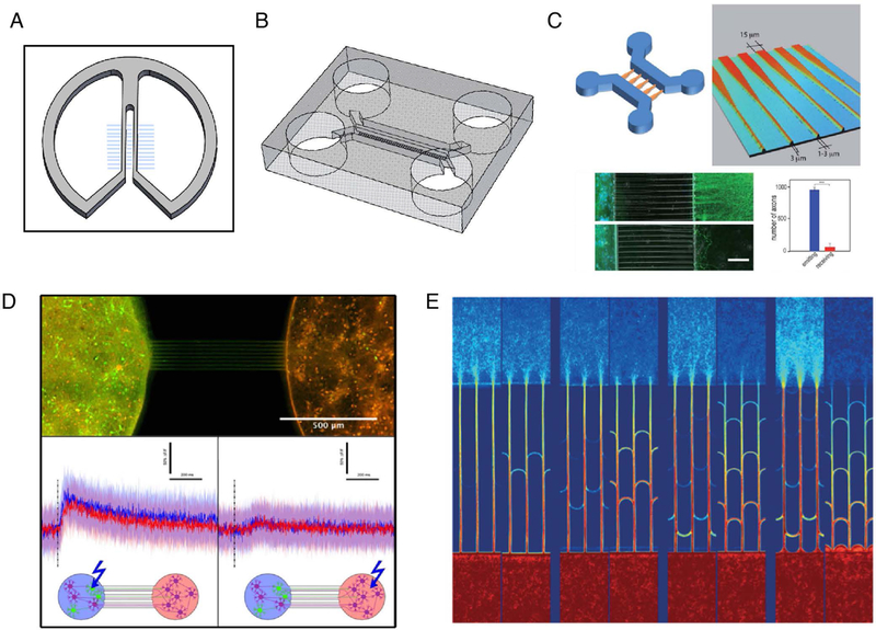
Figure 1.

Tools for compartmentalized systems. A) Campenot chamber (re-drawn from Zweifel et al. (Zweifel et al., 2005) and Millet and Gillette (Millet and Gillette, 2012)). Scratches in collagen surface produce channels which direct axonal outgrowth. B) Compartmentalized device made from PDMS (re-drawn from Taylor et al. (Taylor et al., 2005)). Microchannels (3 μm tall, 10 μm wide) connect two opposing chambers. C) Axon diodes drive axonal outgrowth primarily in one direction. Wider channels (15 μm) provide greater opportunity for neurites to enter whereas thinner channels (3 μm) limit neurite entry. (Reproduced from Peyrin et al. (Peyrin et al., 2011) with slight modifications with permission.) D) Incorporation of optogenetics and calcium imaging in a culture system, coupled with axon diodes. Calcium imaging bursts in response to light seen in both compartments only when Channelrhodopsin-2-transduced neurons were stimulated. (Reproduced with permission under CCBY4.0(https://creativecommons.org/licenses/by/4.0/legalcode) from Renault et al. (Renault et al., 2015)with slight modifications.) E) “Return-to-sender” approach provides directionality between compartments. Axons entering from the “receiver” side encounter bifurcations (arches) which return them back to their original chamber. Axons from the “sender” side do not encounter bifurcations and pass through to the “receiver” side (Reproduced from Renault et al. (Renault et al., 2016) with permission.)
