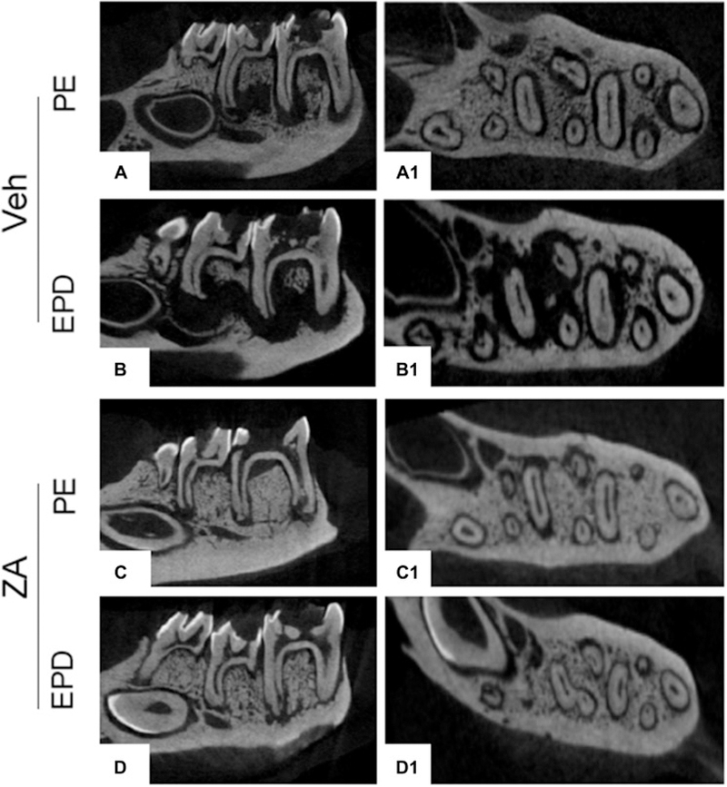
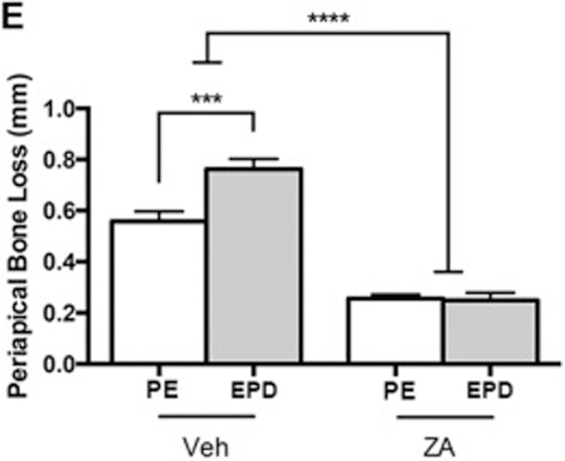
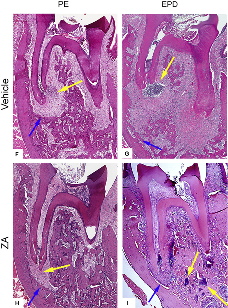
Fig. 2. Radiographic and histologic analysis of pulp exposure & EPD prior to extraction.


µCT assessment of vehicle treated animals in the absence (a, a1) and presence (b, b1) of bacterial inoculation (EPD), and of ZA treated animals in the absence (c, c1) and presence (d, d1) of bacterial inoculation (EPD). (e) Quantification of periapical bone loss. Data represents mean value ± SEM. **** statistical significance, p<0.0001, *** statistical significance, p<0.001, (n=10 per group). Histologic assessment of periapical disease in the absence (f, h) and presence (g, i) of bacterial inoculation (EPD) in vehicle and ZA treated animals, respectively. Blue arrows point to the extent of periapical bone loss. Yellow arrows point to areas of inflammatory infiltrate.
