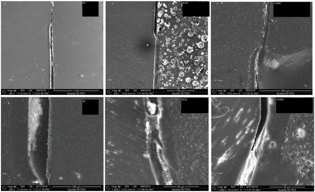
Fig. 4: SEM-analysis.
Micrographs of specimens restored with BC (A & B); BC40 (C & D); and rh25BC (D & E) after 2 weeks of remineralization. Gap formation due to shrinkage was evident in all specimens. The formation of a reaction layer was most obvious in rh25BC, clearly present in BC40, and barely detectable in BC.

